| |
Orthotic Case Study
Flat Feet
Patient
Symptoms
Severe stabbing pain in left foot, 1st interphalangeal joint when walking and during passive movement
Severe pain in right foot, lateral side of sub talar joint when walking
Occasional burning pain right foot, 3rd interspace when walking, particularly noticeable when walking barefoot
| Clinical Observations |
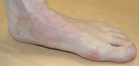
Left Foot - Medial View
Patient Standing
Flat medial longitudinal arch, alternatively called pes planus, flat foot, fallen arch or foot rolling in.
Internally rotated hallux
|
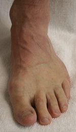
Left Foot - Dorsal View
Patient Standing
Internally rotated hallux
Developing Hallux Valgus
Previously partial nail avulsion medial side of hallux nail - the internal rotation of the hallux encouraged the medial side of the nail to rub against the shoe at propulsion
2nd toe - starting to contract |
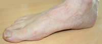
Right Foot - Medial View
Patient Standing |
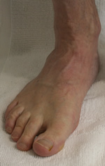
Right Foot - Dorsal View
Patient Standing |
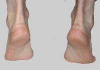
Posterior View
Patient standing on toes
Shows heel inversion suggesting the tibialis posterior muscle is functioning. The heel inverts as the tibialis posterior contracts because it is the main invertor of the foot. |
Ankle Joint Dorsiflexion
Adequate for normal walking |
Treatment
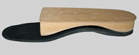
Orthotics (functional insoles to wear inside shoes) provided to control excessive pronation, change walking and standing, deflect pressure and support the foot. |
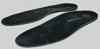
Custom made orthotics provided to help pain |
 |
|
Biomechanical Examination and Gaitscan
To help determine treatment and orthotic prescription |
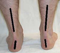
Bisection Lines marked on heel and calf
Patient standing in Relaxed Calcaneal Stance Position (RCSP) i.e. normal standing position
Prominent left and right medial malleoli
RCSP 5 deg valgus left and right |
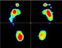
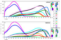
Gaitscan Static Measurement
Shows high pressure areas in red |
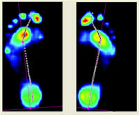
Gaitscan Dynamic 2D photo |
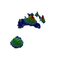
Gaitscan - 3D. left foot |
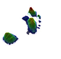
Gaitscan - 3D. right foot |
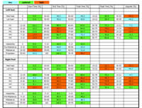
Gaitscan - Report and Measurements |
Follow Up
Pain free at 2 month follow up |
|
|
|
|
Contact Sue Ferguson
Podiatrist
Book an appointment now to help your foot pain (please note I am not accepting new patients)
Tel: 01580 765546
|
| |
| |
|